
Diagnostic Imaging
Baylor Scott & White Medical Center – Trophy Club is pleased to present a comprehensive diagnostic imaging center to the Dallas/Fort Worth area. We are the leading provider of advanced diagnostic imaging technologies with state-of-the-art equipment and highly qualified and caring technologists.
The center is open Monday through Friday from 8:00 a.m. to 5:00 p.m. to serve you. After-hours appointments are also available Monday through Friday, 5:00 p.m. to 11:00 p.m., and Saturdays and Sundays from 8:00 a.m. to 11:00 p.m. We accept walk-ins with a physician’s orders.
Imaging Services at Baylor Scott & White Medical Center – Trophy Club:
CT (Computed Tomography)
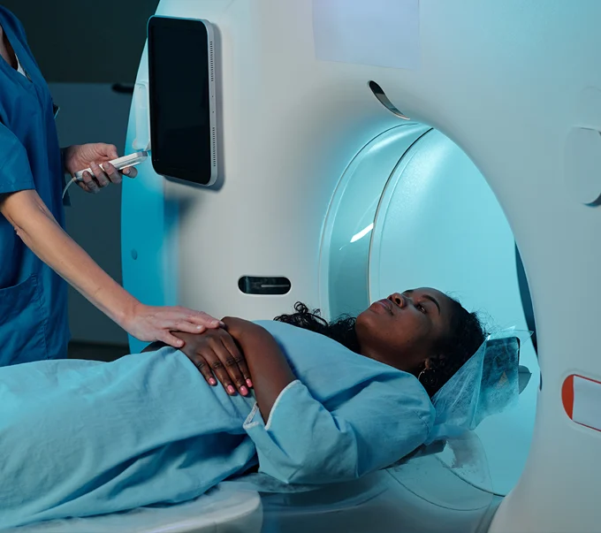
A “CT” or “CAT” scan is used to describe a radiological test known as computed tomography. This test is an innovative way of looking at the inside of the body. The CT scanner is donut-like in shape. The images it produces are cross-sectional-patterned, much like slices of bread. A CT scan can create a multidimensional view of your body by taking a series of such embodiments.
CT makes it possible to diagnose various medical conditions because it sees the inside of the head and body that cannot be seen on regular X-rays.
For many computed tomography examinations, you may be asked to take a unique contrast material (orally, rectally, and/or by injection). Intravenous (IV), oral, and rectal CT contrast liquids are sometimes called “dye.” CT contrast is used to make specific organs, blood vessels, and/or tissue types “stand out” to show the presence of disease or injury.
Radiology/ Fluoroscopy
X-rays are an excellent addition to medicine; they allow doctors to peer into the human body without a surgical procedure. Examining a broken bone with X-rays is much easier and safer than opening a patient up.
Radiography, known to laypeople as “X-ray,” is the oldest and most commonly used type of medical imaging. X-rays are created by passing a small, highly controlled amount of ionizing radiation through a specific human body area, capturing the resultant shadows and reflections on a digital plate.
You may be asked to swallow a radioactive liquid called a barium during a fluoroscopy. This liquid is harmless and shows up in the movie-like sequence of images. This imaging technique is often used to look at the internal organs that play a part in swallowing and digestion, like the esophagus and stomach.
Other X-ray imaging capabilities include gastrointestinal, genitourinary, myelograms, hysterosalpingograms, musculoskeletal studies, arthrograms, and entire spine and bone length exams.
Magnetic Resonance Imaging
Magnetic Resonance Imaging (MRI) is the method of choice for evaluating many different types of injuries and conditions due to its ability to tailor exams to the particular medical question.
Images of unparalleled detail are obtained using a strong magnetic field, particular radio frequencies, and a computer without ionizing radiation.
Ultrasound
Ultrasound imaging is a relatively inexpensive, fast, and radiation-free modality. Ultrasound is excellent for the non-invasive diagnosis of several conditions. Ultrasound is used extensively to show fetal development and to evaluate the kidneys, liver, gallbladder, pancreas, heart, and blood vessels.
Patients often lie down for the sonogram. A clear, water-based conducting gel is applied to the skin over the examined area to help transmit the sound waves. The handheld ultrasound transducer is then moved over the area of interest.
Digital Imaging
Baylor Scott & White Medical Center – Trophy Club employs the highest in technically advanced radiology and digitally enhanced imagery. All images are saved as files and can be sent via a secure website or burned to CD. Images are immediately viewable by physicians on specialized computer screens.